Recently, the teams of Liu Chengbo and Zheng Wei from the Center for Biomedical Optics and Molecular Imaging at Shenzhen Institutes of Advanced Technology, Chinese Academy of Sciences, collaborated and published a research paper titled "Video-rate high-resolution single-pixel nonscanning photoacoustic microscopy" in Biomedical Optics Express, a flagship journal in the field of biomedical optics. The paper reported a high-speed photoacoustic microscopy imaging technology based on a single-pixel non-scanning method. For the first time internationally, it achieved three-dimensional dynamic photoacoustic microscopy imaging at a rate of 30 frames per second, reaching the fastest imaging speed among similar technologies. This paper was selected by the journal as an Editor's Pick highlight article. Assistant Researchers Chen Ningbo, Yu Jia, and Liu Liangjian are the co-first authors of the paper, and Researcher Liu Chengbo and Researcher Zheng Wei are the co-corresponding authors.

Screenshot of the paper when it was published online
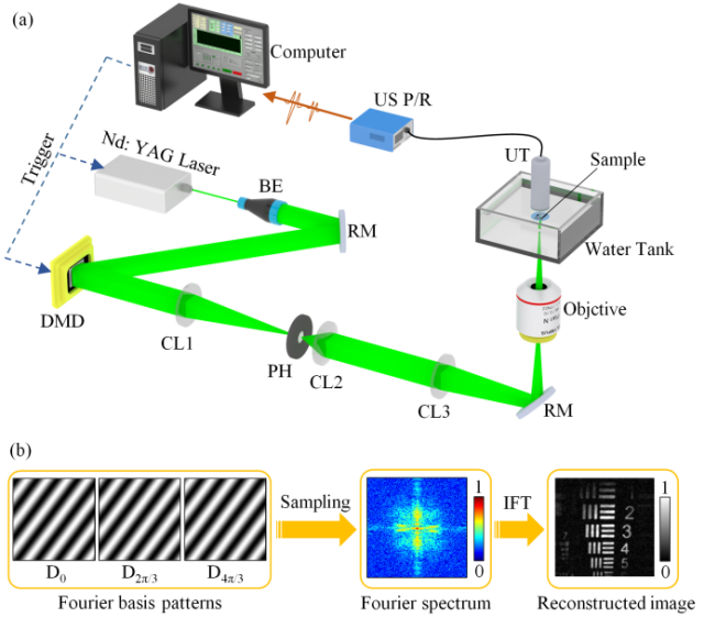
Photoacoustic microscopy imaging has the advantages of high resolution, three-dimensional label-free imaging, etc., and is widely used for the structural and functional imaging of living biological tissues. Traditional photoacoustic microscopy imaging technology mainly relies on point-by-point scanning for three-dimensional image acquisition. Limited by the scanning speed of scanning devices such as stepper motors and optical galvanometers, the imaging speed of traditional photoacoustic microscopy technology is much lower than the video frame rate (30 Hz), making it difficult to meet the needs of monitoring the rapid physiological activities of organisms. To address this issue, the research team proposed a high-speed non-scanning photoacoustic microscopy (SPN-PAM) based on single-pixel imaging technology. This technology uses a high-speed digital micromirror device (DMD) to achieve structured light field illumination of the imaging area. By rapidly modulating the Fourier illumination fringes of the structured light field, the spectral information of the transform domain of the image is obtained, and the rapid reconstruction of the image can be completed by using the inverse Fourier transform of the frequency spectrum.

(a) Diagram of the SPN-PAM imaging system; (b) Principle of SPN-PAM image reconstruction
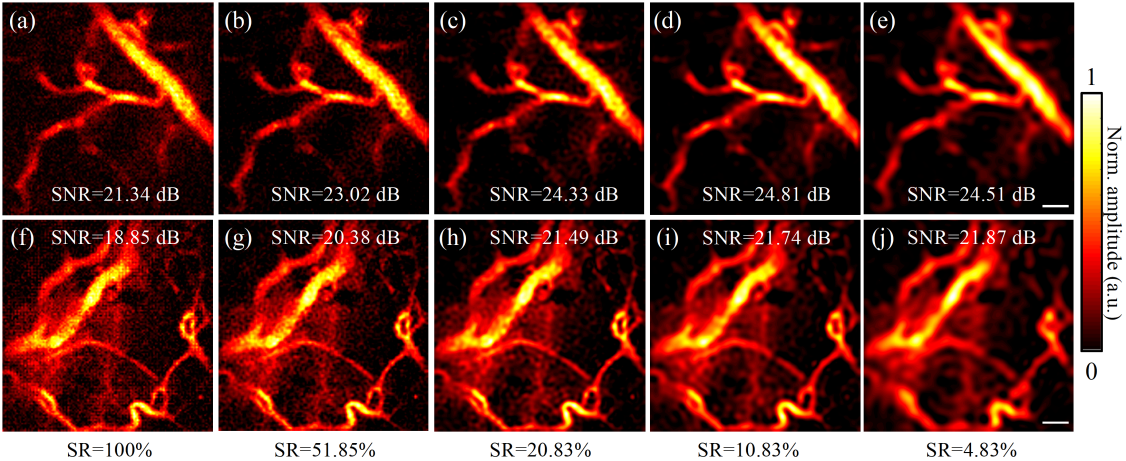
The SPN-PAM technology does not require point-by-point scanning for imaging, overcoming the limitation of scanning devices on the imaging speed. In addition, this technology can make full use of the unique spectral sparse characteristics of the image in the transform domain and perform significant compression sampling of the spectral information. The in vivo imaging results show that under an extremely low sampling rate of 4.86%, this method can still maintain good image resolution and signal-to-noise ratio, while the imaging speed is greatly improved. Based on this, the research team has achieved high-resolution photoacoustic microscopy imaging at the video frame rate (≥30 Hz) for the first time.

Results of SPN-PAM Compressed Imaging of the Microvascular Network in a Living Mouse
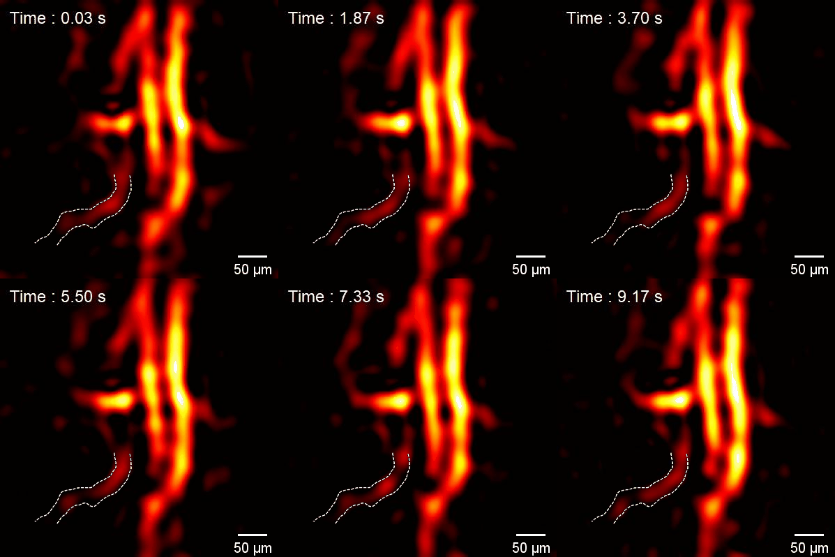
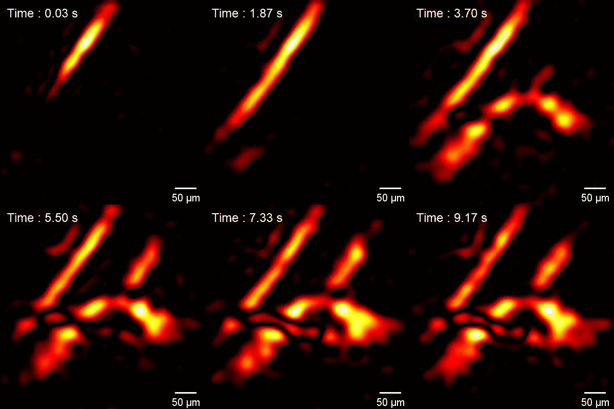
With the advantages of high temporal and spatial resolution, SPN-PAM has achieved dynamic monitoring of the blood flow reperfusion process at the microvascular scale in living small animals at the in vivo level. It has observed the instantaneous changes in blood flow volume and velocity, providing a potentially effective means for the study of hemodynamics and tissue metabolism. At the same time, SPN-PAM compressed imaging can also effectively reduce the laser dose required for high-speed photoacoustic microscopy imaging, enhancing the imaging safety and offering the possibility for the further clinical translation of this technology.


Dynamic monitoring of microvascular blood flow (left) and the blood flow reperfusion process (right) in a living mouse
This work was supported by projects such as the Key R&D Program of the Ministry of Science and Technology, the National Natural Science Foundation of China, the Chinese Academy of Sciences, and the Key Laboratory of Guangdong Province.