Recently, the Biomedical Optics and Molecular Imaging Laboratory at the Shenzhen Institutes of Advanced Technology (SIAT), Chinese Academy of Sciences, has achieved a significant breakthrough in optical-resolution photoacoustic microscopy (OR-PAM). The team successfully developed the world’s first label-free, high-resolution (5 μm) quantitative imaging technique for visualizing the vasculature of the anterior eye segment in live animals. This innovative method not only captures detailed microvascular networks but also enables fully automated vessel enhancement, extraction, and quantitative analysis. It holds great promise as a novel tool for high-curvature superficial vascular imaging and quantitative assessment, with potential applications in diagnosing human ocular vascular diseases.
The research findings, titled "Three-dimensional Hessian matrix-based quantitative vascular imaging of rat iris with optical-resolution photoacoustic microscopy in vivo," were published in the Journal of Biomedical Optics—a leading journal in biomedical optics—and featured as the cover article for its May 2018 issue.
Imaging and quantitative analysis of the iris microvasculature are crucial for diagnosing and evaluating ocular diseases. Photoacoustic imaging offers exceptional sensitivity to endogenous hemoglobin, providing an efficient, label-free approach for visualizing iris vasculature. To advance this technology, the team developed a vascular quantification algorithm based on a 3D Hessian matrix, enabling precise extraction of microvascular data. Integrated with their self-developed OR-PAM system, this algorithm successfully generated 3D images and quantitative metrics of the iris microvasculature.
For the first time, the study achieved label-free imaging of the microvascular architecture in live rat irises, accurately quantifying key parameters such as vessel diameter, density, and tortuosity. This fully automated vascular extraction and quantification technique enhances OR-PAM’s diagnostic potential for ocular vascular disorders and may soon be adapted for clinical use in human eye disease diagnosis.
The study was conducted under the joint supervision of Dr. Chengbo Liu (Associate Researcher at SIAT) and Prof. Zhicheng Liu (Capital Medical University), with Ph.D. candidate Huangxuan Zhao as the lead researcher. The SIAT team maintains a long-term collaboration with Capital Medical University, jointly advancing photoacoustic imaging technology for early diagnosis of ocular vascular diseases.
This work was supported by grants from the National Natural Science Foundation of China, the National Basic Research Program (973 Plan), the Beijing Natural Science Foundation, and the Shenzhen Basic Research Program.
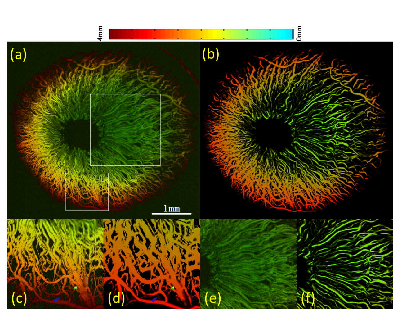
(a) Custom OR-PAM displays the depth-encoded map of rat iris vasculature; (b) Depth-encoded map of rat iris vasculature after algorithmic enhancement; (c) and (d) show comparative views of vascular signals in the focal region before and after enhancement, respectively; (e) and (f) present comparative views of vascular signals in distant out-of-focus regions before and after enhancement, respectively.
